The Bell Brothers and the Art of Surgical Anatomy
Dates: Feb. 7 to Sept. 30, 2024
Contact: ghsl-special
At the turn of the 19th century, Scottish brothers John and Charles Bell set out to reform medical education and, by extension, medicine. Through their work as teachers of surgery and anatomy and as authors of medical textbooks, they aimed to provide students with an experiential education that they could apply to their future careers as physicians and surgeons.
What made the Bells' work especially distinctive was their artistic skills, which they used to illustrate their own books. Combining their knowledge of anatomy, their real-world experience as surgeons, and their clear, realistic artistic styles, the Bell brothers presented anatomy and surgery from a practical perspective, encouraging their students to apply this knowledge to their own practice.
The Bells and their World
Brothers John and Charles Bell were born into an accomplished Edinburgh family. Their father, Reverend William Bell, died when they were young, leaving their mother, Margaret, to raise them.
John and Charles each had successful careers as teachers of surgery and anatomy, as practicing surgeons, and as artists who illustrated their own medical works. Explore this exhibit to learn how their artistic work contributed to their broader goals of improving the teaching and practice of medicine.
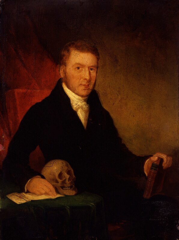 John Bell
- Born in 1763 in Edinburgh, Scotland
- MD from the University of Edinburgh, 1779
- Fellow of the Royal College of Surgeons of Edinburgh, 1786
- Started his own school of anatomy in Edinburgh
- Had the most successful surgical practice in Scotland
- Regarded as the father of applied surgical anatomy
- Died in 1820 in Rome, Italy
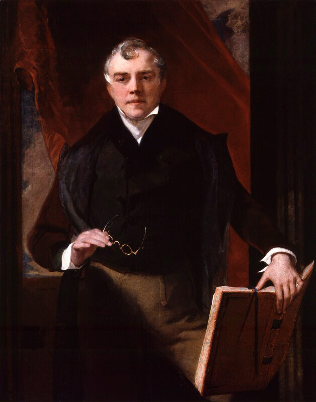 Charles Bell
- Born in 1774 in Edinburgh, Scotland
- Studied medicine at the University of Edinburgh
- Fellow of the Royal College of Surgeons of Edinburgh, 1799
- Taught surgery and anatomy in London
- Involved in founding King's College London and University College London's medical school
- Known for studying the nervous system and for describing Bell's palsy, a type of facial paralysis
- Died in 1842 in Hallow, England
The Scottish Enlightenment
![Painting titled The Parliament Close [Edinburgh] by John Le Conte after David Wilkie, 1844.](../../../../images/exhibits/bell-brothers/parliament.jpg)
Scotland was one of the centers of the Enlightenment, the intellectual and philosophical movement that swept through Western Europe in the 18th century. Its proponents valued human reason and skepticism over dogmatic religious tradition and emphasized the pursuit of empirical scientific knowledge to benefit the individual and society.
Already a center of learning, with five universities compared to England's two, Scotland—and particularly Edinburgh—had the right intellectual environment to generate new ideas across all areas of knowledge, including medicine. Many of the great physicians and surgeons of the time, like John and Charles Bell, were Scottish, including brothers John and William Hunter, father and son Alexander Monro Primus and Alexander Monro Secundus, and Benjamin Bell (no relation).
The State of Medicine
The Enlightenment's focus on experimentation and empiricism in science extended to medicine, which saw numerous advances in the periods preceding and during the Bells' lifetimes. Physicians conducted rigorous observations of the human body to better understand its functioning, and they collected specimens in museums to enhance the study of the body and its diseases, injuries and disabilities.
Another pair of Scottish brothers, William and John Hunter, had significant impacts on anatomy and surgery in the mid-18th century. William opened his own anatomy school in Great Windmill Street in London, where he emphasized the importance of dissection; Charles Bell would later teach there after its incorporation into King's College London. John is considered the father of scientific surgery for bringing the Enlightenment principles of careful observation and the scientific method to surgical practice.
The State of Medical Education
Progress brought about by the Enlightenment also affected medical education. The focus on empiricism and experimentation in science and medicine created a demand for a more formal and rigorous education. Even so, educational standards still varied: many surgeons and apothecaries (who could dispense medications and would later become general practitioners) had no university education and instead trained as apprentices, with some instruction at hospitals or private schools, such as those run by the Bells. It wasn't until 1858 that strict standards were set for the education and qualification of doctors in Britain.
Edinburgh was a major center of medical education in Britain; the first medical school in the English-speaking world had been founded at the University of Edinburgh in 1726. As the city's reputation grew, more students came there to attend its many public and private medical schools, including John Bell's anatomy school in what was called Surgeons' Square.
The Bells as Artists
Because of their father's early death, the Bells' mother, Margaret, had a significant influence on their early lives, particularly on the youngest son, Charles. An accomplished artist herself, she passed on a talent for drawing to her sons and even paid for Charles to take lessons in painting and drawing from the director of the Edinburgh Academy of Arts. Both brothers used these skills to illustrate their own medical texts.
Medical writers who illustrated their works were rare. Those who didn’t have to hire artists who often had less knowledge of anatomy, risking inaccuracies. The Bells also taught themselves etching to avoid further errors when transferring their drawings to the metal plates used for printing.
Even in... our best anatomical figures, we see a continual struggle between the anatomist and the painter; one striving for elegance of form, the other insisting on accuracy of representation.
John Bell, preface to Engravings, Explaining the Anatomy of the Bones, Muscles, and Joints.
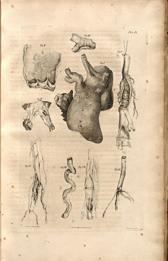
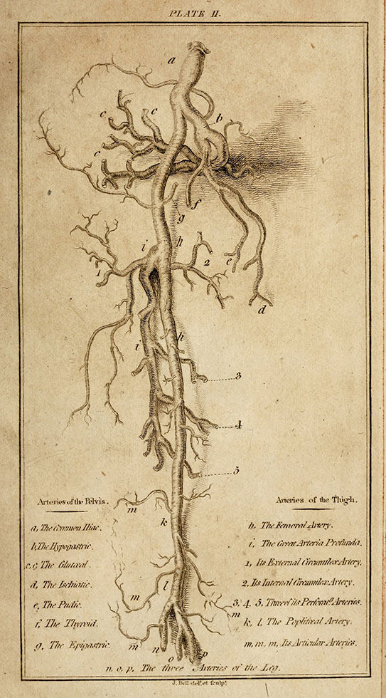
The two plates on display here show images both drawn and etched by each of the Bells. By performing both parts of the illustration process themselves, they ensured the accuracy of the anatomical details.
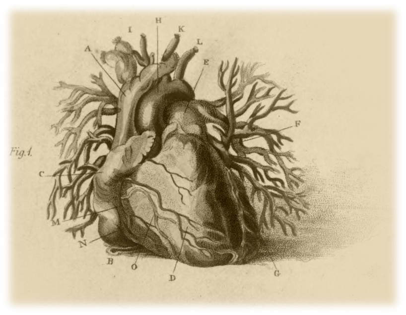
Above left: Plate 9. Illustration of the heart, drawn and etched by Charles Bell. Charles Bell. A System of Dissections, Explaining the Anatomy of the Human Body. Edinburgh: Printed for Mundell and Son, 1798-1803.
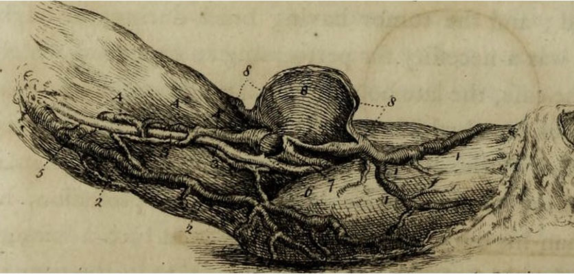
Above right: Plate 2. Illustration of the arteries of the pelvis and the thigh, drawn and etched by John Bell. John Bell. Discourses on the Nature and Cure of Wounds. Edinburgh: Bell & Bradfute, 1795.
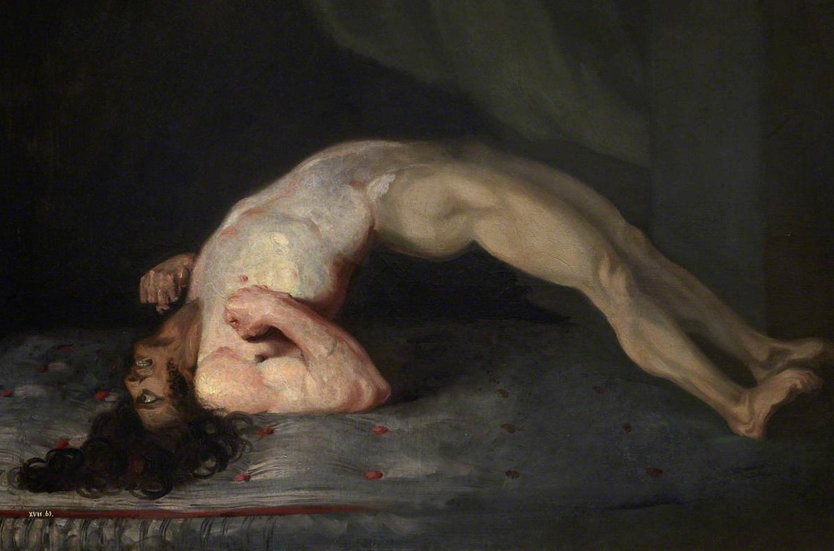
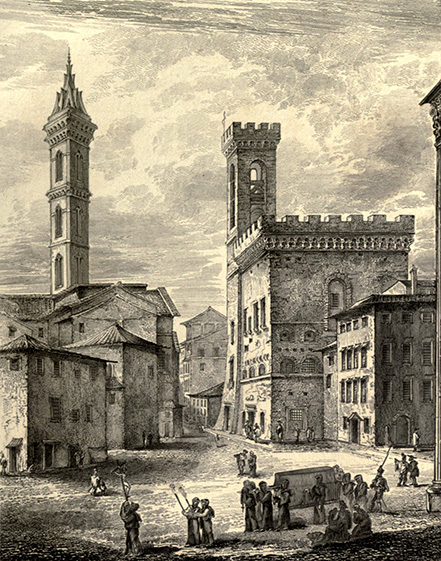
The Bells' artistry extended beyond their anatomical drawings. On display are additional examples of their talent: Charles's painting of a soldier suffering from tetanus caused by a gunshot wound and an engraving of John's architectural illustration in Florence, Italy.
Above left: The Wounded following the Battle of Corunna: Tetanus Following Gunshot Wounds. Painted by Charles Bell, 1809. From the Royal College of Surgeons of Edinburgh collection.
Above right: "View of Badia and Podesta." Drawn by John Bell and engraved by W.H. Lizars. John Bell. Observations on Italy. Edinburgh: William Blackwood, 1825.
Artistic Style
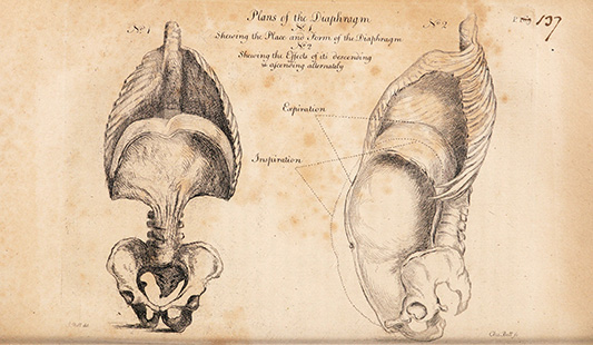
John and Charles shared goals regarding the style of their anatomical artwork: clarity and realism. The Bells favored plainness over flourish: simple, clear images with straightforward descriptions would help students learn more effectively than fanciful illustrations with Latin text.
Right: John omits much of the thorax and abdomen to focus the viewer’s attention on how the diaphragm moves in concert with the lungs, ribcage, and abdominal muscles during respiration.
I have ventured...to make every description as simple as may be,—using no hard words, but the pure names; choosing rather that my book should be plainly understood, than admired as a piece of unintelligible profound anatomy.
John Bell, Engravings, Explaining the Anatomy of the Bones, Muscles, and Joints, xxii
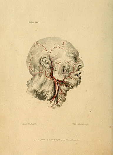 Left: Plate 3. Drawn by Charles Bell, etched by T. Medland. Charles Bell and John Bell. Engravings of the Arteries, Illustrating the Second Volume of the Anatomy of the Human Body. London: Longman and Rees, 1801.
[T]he design of this book of Plates is to present to the student, at one glance, the general distribution of the vessels, and to fix them in his memory in a way which no description can accomplish.
Charles Bell, preface to Engravings of the Arteries
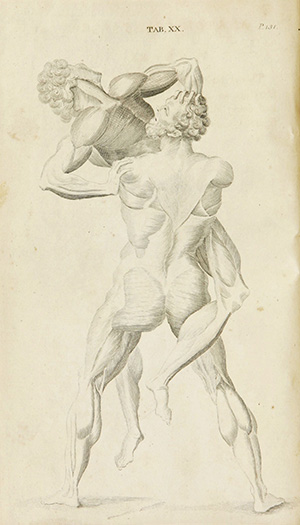 Right:
This pre-Enlightenment book was the textbook of the English medical schools for many years, running through 13 editions. For this plate, Cheselden omitted an explanation and a key because he believed that alphabet labels would spoil the image's beauty. He sacrifices clarity and information to present his artist’s anatomical version of a famous classical statue of Hercules.
Plate 20. William Cheselden. Anatomical Tables of the Human Body. Boston: printed by Manning and Loring, for David West, 1796.
Realism at the Individual Level
The Bell brothers were far from the first writers to strive for realistic depictions of anatomy in their work. For centuries, anatomists prided themselves on presenting images made from direct observation. Most anatomists examined many bodies and then created composite images that depicted an idealized anatomy.
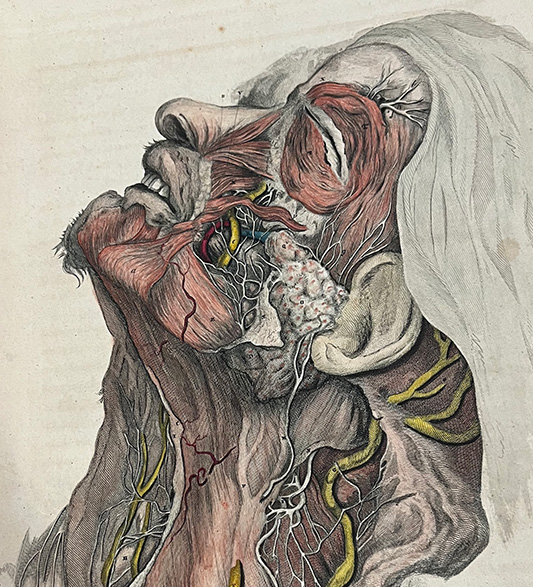
Charles also examined many bodies for his illustrations, but did so to select the best one to depict the issue. Elements that could easily be omitted or obscured—facial hair, clothing—are intentionally included, signaling that this is an individual human being as well as an individual case study. Charles portrayed nature exactly as it is found and then used the text to indicate what characteristics could be universalized across bodies
Similarly, John described general anatomical knowledge in his text while depicting a highly specific example, indicating what an image would and would not depict, including what a surgeon might encounter in real life.
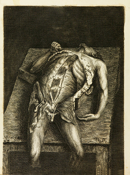
Above left: John presents this body exactly as a cadaver would appear in a dissection theater: propped on a box to provide spinal curvature so that students in the upper tiers could see the demonstrator performing the dissection.
Book 2, plate 9. Muscles. Drawn by John Bell and engraved by John Beugo. John Bell. Engravings, Explaining the Anatomy of the Bones, Muscles, and Joints. Edinburgh: Printed by John Paterson, for Bell and Bradfute, etc., etc., 1794.
Above center: Elements that could easily be omitted or obscured—facial hair, a head covering, the undissected ear, nose, and lips—are intentionally included, signaling that this is an individual human being as well as an individual case study.
Plate 5. Drawn by Charles Bell and engraved by D. Lizars. Charles Bell. A System of Dissections. Vol. 2. Edinburgh: printed for Mundell and Son, 1798-1803.
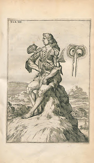
Above right: In this anatomy, many of the figures are portrayed helpfully holding layers of skin or muscle to isolate the described anatomy for the student. These whimsical illustrations are intended to depict muscles as they would appear in a living body, but they do not necessarily reflect what a physician would encounter in practice. Conversely, John and Charles seek to create realistic depictions of the human body, whether it is a cadaver awaiting dissection or a living body in need of surgery.
Plate 12. John Browne. Myographia Nova. Lugduni Batavorum: apud Jacobum Mouckee, 1687.
Same Goals, Different Execution
While Charles and John shared similar objectives for their images, they differed in how they treated their morbid subject matter. Charles sought to present reality while also maintaining the human dignity of his subject. He employs an elegant, fine-line style to depict bodies accurately and precisely, while also producing an aesthetically pleasing image. He tended to include personal characteristics to retain the element of humanity in a cadaver, or to isolate a body part in abstraction to present a detached view.
The bodies in John's images are portrayed in a harsher light. He chose to present the intense reality of the anatomical theater—a dark, crude, cruel room evocative of a slaughterhouse—so the student will be prepared when they do witness the dissection of a human body.
Anatomical Education
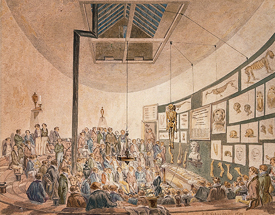
John and Charles Bell both taught anatomy and surgery during their careers, John in Edinburgh and Charles in London. Each set up his own private school to provide anatomical instruction that would give students an education grounded in practical experience.
At right: This watercolor shows the classroom in the anatomy school started by William Hunter in London’s Great Windmill Street. Charles Bell taught here from 1811 to 1824. Although this image was painted in 1839, it illustrates the variety of visual and tactile methods used to teach anatomy.
Like many of the leading medical educators of the time, the Bells wrote their own textbooks on surgery and anatomy. They used the illustrations in their books, which they drew themselves, to supplement the texts and their classroom instruction. They believed that illustrations were an important tool to help students absorb the information they learned.
Drawings are a kind of notes, too, more easily consulted; and bring to the mind, in a more lively manner, all that was associated in our first studies.
—Charles Bell, preface to Engravings of the Arteries, Illustrating the Second Volume of the Anatomy of the Human Body.
The illustrations that the Bells created for their textbooks were part of a multimodal pedagogical program that used visual and tactile examples to teach anatomy, which could then be applied to surgery. These examples included wax models, preserved specimens, paintings and sculptures. Students viewed these and created their own, to train both their eyes and their hands.
A book of anatomy without [plates] seemed to me no better than a book of geography without its maps... like teaching mathematics without diagrams.
—John Bell, preface to Engravings, Explaining the Anatomy of the Bones, Muscles, and Joints.
Most important was dissection of a human body. Students' anatomy textbooks often served as guides for dissection, providing detailed directions and explanations of what they would observe. Despite the Bells' emphasis on practical experience, however, dissection at the turn of the 19th century still followed a pattern set hundreds of years earlier: students would watch as a demonstrator performed the dissection and the professor narrated what they were seeing. Most students wouldn't have had the opportunity to dissect bodies themselves until later in the 19th century, after new legislation permitted the procurement of more bodies for medical schools.
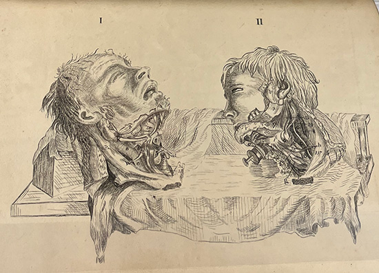
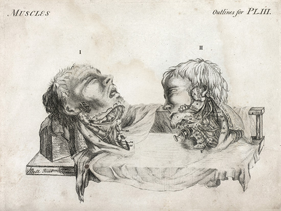
Above: A common exercise and method of study for medical students in the 18th and 19th centuries was to copy the text and illustrations from their textbooks. This helped to train their hands as well as their eyes. The notebook at left was copied by a man named John Brettell in 1801 from John Bell’s Engravings, Explaining the Anatomy of the Bones, Muscles, and Joints. At right is the corresponding illustration from Bell’s book.
Book 2, plate 3. Drawn and engraved by John Bell. John Bell. Engravings, Explaining the Anatomy of the Bones, Muscles, and Joints. Edinburgh: Printed by John Paterson, for Bell and Bradfute etc., etc., 1794.
Grave Robbing
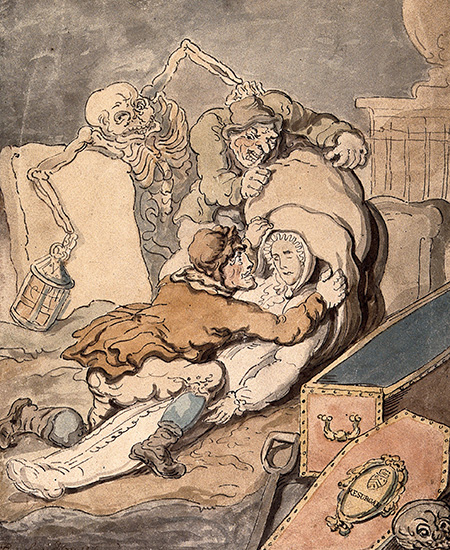 Although dissection was an increasingly important part of medical students' training by the early 1800s, obtaining bodies had always been difficult because of laws prohibiting human dissection. Earlier legal reforms allowed specific institutions to procure a limited number of bodies, but it was often insufficient to meet the demands of the growing number of medical schools. The practice of grave robbing became common in the 1800s to provide schools with the bodies they desperately needed to teach their students.
The most famous "body snatchers," Burke and Hare, who resorted to murder to supply bodies to an anatomist, operated in Edinburgh. While we can't know for sure if the Bells used illegally-obtained bodies in their schools, it wouldn't be surprising if they participated in the illicit trade to ensure a complete anatomical education for their students.
At right: Watercolor drawing of body snatchers by Thomas Rowlandson, 1755.
Anatomical Education Today
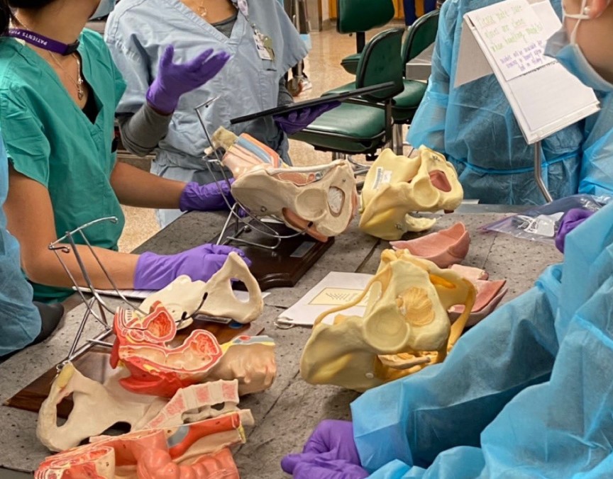
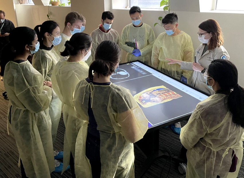
Many aspects of medical education have changed in the 200 years since John and Charles Bell were active, but the multimodal approach to teaching anatomy remains important for developing a well-rounded understanding of the structure and function of the human body.
Advances in technology, from the invention of photography and motion pictures to the use of E-books and virtual reality, have enhanced students' visual experience of human anatomy, but the practical, hands-on experience of dissection remains foundational in modern medical education. Even so, some medical schools have begun moving away from using donated bodies, which are expensive to maintain and still difficult to procure in sufficient numbers, and are instead using interactive three-dimensional software to better represent the living human body. Who can say what the landscape of anatomical education will look like in another 200 years?
These items represent some of the different types of learning tools that were used in medical education throughout the 20th century:
- Wax embryo model (ca. 1880)
- Bailliere’s Synthetic Anatomy (1928)
- Viewmaster and Stereoscopic Atlas of Human Anatomy (1954)
Pictured here are just some of the types of visual and tactile learning tools that students use to study anatomy at Northwestern University Feinberg School of Medicine:
- Plastic models
- Touchscreen table using Visible Human Dissector
- 3D mixed reality software

The Birth of Surgical Anatomy
From 1720 to 1846, anatomical education in Edinburgh was dominated by the Monro family. John Bell took issue with their invincible monopoly, noting that they were not practicing surgeons and therefore could not teach surgical anatomy with any expertise.
The Bells believed that anatomy courses should not solely focus on a student’s mastery of anatomical knowledge for its own sake. Instead, the practical application of anatomy as it relates to surgery—that is, surgical anatomy—should be the ultimate goal. John realized this aim when he opened the first school of surgical anatomy in 1790.
To ally anatomy more closely with surgery, to connect the art more intimately with the science from which it flows, seemed to me a task as yet imperfectly accomplished and yet of the very highest importance.
—John Bell, preface to The Principles of Surgery
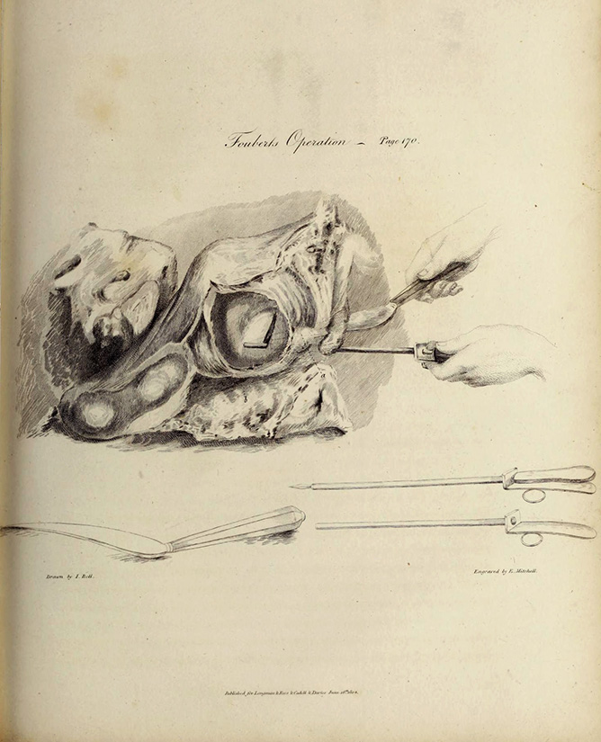 At right: Instead of simply recommending the best method to remove bladder stones, John reviews the evolution of the procedure and details in simple terms the dangers and imprecision of past methods. As an empiricist, he presents all the evidence—including images—so that readers know that his conclusions and recommendations are grounded in personal experience. For his discussion of Foubert’s Operation for lateral lithotomy, he illustrates the specialized instruments both in use and as diagrams.
Plate facing page 170. Fouberts Operation. Drawn by John Bell and engraved by E. Mitchell. John Bell. The Principles of Surgery: as They Relate to Wounds, Ulcers and Fistulas, etc. Vol. 2, pt. 1. London: Longman, 1815.
Theory vs. Practice
Amputation was a common operation during wartime and boasted only a 25% success rate when performed in a controlled, though unsterilized, hospital setting. It was far less successful in the field. Charles operated on wounded soldiers after the battles of Corunna and Waterloo during the Napoleonic Wars (1803-1815), and this illustration may depict an operation he performed or witnessed.
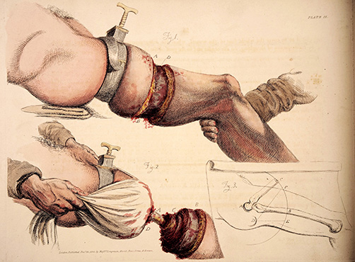
Above: This engraving depicts the grim realities of a surgical amputation above the knee. Charles clearly explains his process in plain language, articulating why certain steps are necessary or beneficial based on his experience. The hand-coloring conveys the realities of surgery: the bloodiness of the incisions and the pallor of the skin both provide useful information for the reader and serve as an artistic and horrific contrast.
Plate 9. No artists are credited for this illustration, but other plates in the book are drawn by Charles Bell and etched by Thomas Landseer. Charles Bell. Illustrations of the Great Operations of Surgery: Trepan, Hernia, Amputation, Aneurism, and Lithotomy. London: Longman, Hurst, Rees, Orme, and Brown, Paternoster-Row; by A. and R. Spottiswoode, Printers-Street, 1821.
Anatomical Traditions: Perfection and Objectivity
Fifty years before John Bell wrote his anatomy, Dutch anatomist Bernhard Albinus published an enormous atlas that he hoped would set a new standard for accurate and beautiful anatomical art. Like the Bells, Albinus studied many different cadavers in preparation for his book, but did so to calculate proportions for an "ideal" human figure. He believed symmetry and idealization reflected the balance of a healthy body.
At the same time, Albinus valued objectivity and precision. Not an artist himself, he developed a multistep grid system for his illustrator to draw life-size images that could be meticulously scaled down and then engraved. While the figures were drawn from life—that is, from corpses or living people—Albinus made his artist continually alter the drawings so his ideal proportions were portrayed over individual details.
Authority and Control
Though Albinus hired Jan Wandelaar, an accomplished scientific artist, for this lengthy project, he emphasizes that he was directly involved in every part of Wandelaar's illustration process, going so far as to dismiss the artist's contribution, saying Wandelaar was "entirely ruled by me, as if he was a tool in my hands, and I made the figures myself."¹
Albinus emphasizes this point to reassure the reader that they can trust the illustrations: the images are rendered by an artist, but it is the anatomist himself who ensures accuracy in every respect. Because the Bells were so often responsible for their own artwork, the question of authority is not pertinent.
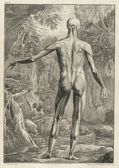
Above left: This idealized representation of the human body highlights not only a difference from the Bells — especially John—in style but also their different goals. While Albinus’ illustrations are steeped in theory and philosophy, the Bells are interested in the practical application of their material.
Table 5. Drawn by Jan Wandelaar and engraved by S.F. Ravenet. Bernhard Siegfried Albinus. Tabulae sceleti et musculorum corporis humani. Londini: Typis H. Woodfall, impensis Johannis et Pauli Knapton, 1749.
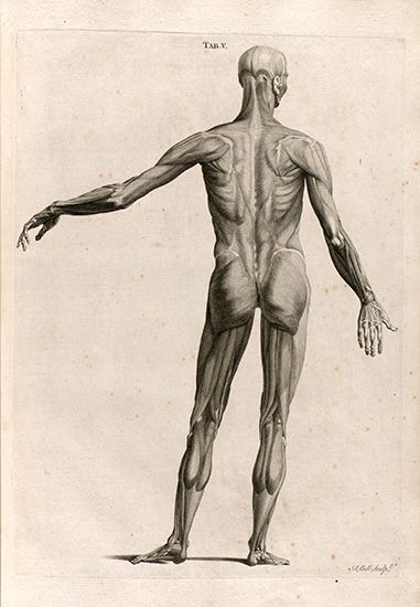
Above right: This faithful copy of Jan Wandelaar’s original figure was replicated for a smaller edition of Albinus’ atlas translated into English. The engraver has notably omitted the natural background. In his first book, Engravings, Explaining the Anatomy of the Bones, Muscles, and Joints, John remarked that the different textures of bone, muscle, and fat are lost in this style and have been “rounded down to the smoothness of ivory.” Students learning from these illustrations will be confounded at a dissection since “the drawings and the subject never can be directly compared (vii-x).”
Table 5. Engraved by Andrew Bell (no relation). Bernhard Siegfried Albinus. Tables of the skeleton and muscles of the human body. Edinburgh: Printed by Balfour & Smellie, for Andrew Bell, engraver, 1777.
Endnote
1. Albinus, Bernhard Siegfried. The Explanation of Albinus’s Anatomical Figures of the Human Skeleton and Muscles. With an Historical Account of the Work. Translated from the Latin (London: John and Paul Knapton, 1754), xi–xii.
 John and Charles Bell sought to publish books with simple text and realistic illustrations in order to make learning anatomy and surgical methods straightforward for students and new physicians.
The Bells employed their artistic skills to maintain control over their illustrations, thereby creating unique styles. They paired their books with other visual, textual, and tactile aids to develop a pedagogical system designed to improve students’ comprehension, retention, and application of anatomy.
In this way, the Bells established themselves as important figures in the history of anatomy and surgery, not through ostentatious, idealized, or extravagant folios, but through their stated goal of teaching young surgeons the practical application of anatomical knowledge via their affordable, straightforward guides.
Credits
Curated and designed by Katie Lattal, MA, Special Collections Librarian, and Emma Florio, MLIS, Archives & Research Specialist.
Bibliography
Primary Resources
- Albinus, Bernhard Siegfried, Andrew Bell, and Jan Wandelaar. Tables of the Skeleton and Muscles of the Human Body. Edinburgh: Printed by Balfour & Smellie, for Andrew Bell, engraver, 1777.
- Bell, Charles. A System of Dissections: Explaining the Anatomy of the Human Body, the Manner of Displaying the Parts, and Their Varieties in Disease. Edinburgh: printed for Mundell and Son, 1799.
- ———. A System of Operative Surgery: Founded on the Basis of Anatomy. 2d ed. London: Longman, Hurst, Rees, Orme, and Brown, 1814.
- Bell, Charles, and John Bell. Engravings of the Arteries, Illustrating the Second Volume of the Anatomy of the Human Body. London: Longman, and Rees, 1801.
- Bell, Charles, and Thomas Landseer. Illustrations of the Great Operations of Surgery: Trepan, Hernia, Amputation, Aneurism, and Lithotomy. First ed. London: Longman, Hurst, Rees, Orme, and Brown, Paternoster-Row; by A. and R. Spottiswoode, Printers-Street, 1821.
- Bell, John. Discourses on the Nature and Cure of Wounds. Edinburgh: Bell & Bradfute [etc., etc.], 1795.
- ———. Engravings, Explaining the Anatomy of the Bones, Muscles, and Joints. Edinburgh: Printed by John Paterson, for Bell and Bradfute [etc., etc.], 1794.
- ———. The Principles of Surgery. London: Longman, 1815.
- Bell, John, and Charles Bell. The Anatomy of the Human Body. Edinburgh: Printed for Cadell and Davies, London, and G. Mudie and son, Edinburgh, 1797.
- Brettell, John, and John Bell. Drawings Copied from the Engravings of John Bell Explaining the Anatomy of the Bones, Muscles and Joints. Unpublished manuscript, 1801.
- Browne, John, Charles Scarburgh, Giulio Cesare Casseri, and William Molins. Myographia Nova. Lugduni Batavorum: apud Jacobum Mouckee, 1687.
- Cheselden, William, William Manning, James Loring, and David West. Anatomical Tables of the Human Body. Boston: Printed by Manning and Loring, for David West, 1796.
Secondary Resources
- Berkowitz, Carin. "The Beauty of Anatomy: Visual Displays and Surgical Education in Early-Nineteenth-Century London." Bulletin of the History of Medicine 85, no. 2 (2011): 248-78. https://doi.org/10.1353/bhm.2011.0030.
- ———. "Systems of Display: The Making of Anatomical Knowledge in Enlightenment Britain." The British Journal for the History of Science 46, no. 3 (2013): 359-87. https://doi.org/10.1017/S0007087412000787.
- ———. Charles Bell and the Anatomy of Reform. Chicago: The University of Chicago Press, 2015.
- ———. "The Illustrious Anatomist: Authorship, Patronage, and Illustrative Style in Anatomy Folios, 1700–1840." Bulletin of the History of Medicine 89, no. 2 (2015): 171-208. https://doi.org/10.1353/bhm.2015.0028.
- Clarke, Edwin. "History of British Medical Education." British Journal of Medical Education 1, no. 1 (1966): 7-15. https://doi.org/10.1111/j.1365-2923.1966.tb02062.
- "Charles Bell." In Surgeons Database, The Royal College of Surgeons of Edinburgh. https://archiveandlibrary.rcsed.ac.uk/surgeon/3769279-charles-bell.
- Fulton, John F. "History of Medical Education." British Medical Journal 2, no. 4834 (1953): 457-61. https://doi.org/10.1136/bmj.2.4834.457.
- Gardner-Thorpe, Christopher. "John Bell (1763–1820): Brother Artist and Anatomist." Progress in Brain Research 203 (2013): 163-83. https://doi.org/10.1016/B978-0-444-62730-8.00006-2.
- Gholipour, Bahar. "Disappearing Bodies." Scientific American (2019): 12-15.
- The Huntington Library. “Tables of the Skeleton and Muscles of the Human Body: An Exceptional Atlas." Learning Resources. Engaging with Nature: Art, Science, and Inquiry, accessed January 10, 2024, https://huntington.org/educators/learning-resources/spotlight/tables-skeleton-and-muscles-human-body.
- "John Bell." In Surgeons Database, The Royal College of Surgeons of Edinburgh. https://archiveandlibrary.rcsed.ac.uk/surgeon/3769283-john-bell.
- Kaufman, M.H. "John Bell (1763-1820), the 'Father' of Surgical Anatomy." Journal of Medical Biography 13, no. 2 (May 2005): 73-81.
- Kemp, Martin. "Style and Non-Style in Anatomical Illustration: from Renaissance Humanism to Henry Gray." Journal of Anatomy 216, no. 2 (2010): 192-208. https://doi.org/10.1111/j.1469-7580.2009.01181.
- Loudon, I. S. L. "Sir Charles Bell and the Anatomy of Expression." British Medical Journal (Clinical research ed.) 285, no. 6357 (1982): 1794-96. https://doi.org/10.1136/bmj.285.6357.1794.
- Loukas, M., M. Akiyama, M. M. Shoja, B. Yalçin, R. S. Tubbs, and A. A. Cohen-Gadol. "John Browne (1642-1702): Anatomist and Plagiarist." Clinical Anatomy 23, no. 1 (Jan 2010): 1-7. https://doi.org/10.1002/ca.20899.
- Morrell, J. B. "The University of Edinburgh in the Late Eighteenth Century: Its Scientific Eminence and Academic Structure." Isis 62, no. 2 (1971): 158-71. http://www.jstor.org/stable/229239.
- Neher, Allister. "John Bell (1763–1820), Anatomist and Art Theorist." The British Art Journal 21, no. 3 (2020): 62-71. https://www.jstor.org/stable/48693110.
- Pearce, J.M.S. "Sir Charles Bell (1774-1842)." Journal of the Royal Society of Medicine 86 (1993): 913-13. https://doi.org/10.1136/jnnp.56.8.913.
- Risse, Guenter B. "Medicine in the Age of Enlightenment." In Medicine in Society: Historical Essays, edited by Andrew Wear. Cambridge: Cambridge University Press, 1992.
- Struthers, John. "Historical Sketch of the Edinburgh Anatomical School." Edinburgh Medical Journal 12, no. 5 (Nov 1866): 431-57.
- Sutherland, Stewart. "Scottish Enlightenment." In Encyclopedia Britannica. https://www.britannica.com/event/Scottish-Enlightenment.
- Zimmerman, Leo M. "Surgeons and the Rise of Clinical Teaching in England." Bulletin of the History of Medicine 37, no. 2 (1963): 167-78. http://www.jstor.org/stable/44446917.